


上海药物所徐华强课题组多巴胺受体研究成果荣登Cell封面
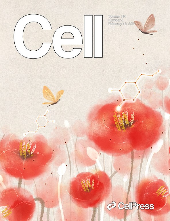
2021年2月11日,上海药物所徐华强课题组联合国内外多家单位于国际顶级期刊Cell以长文形式在线发表了题为Structural insights into the human D1 and D2 dopamine receptor signaling complexes的研究论文1,文章解析了在包括抗帕金森氏病药物溴隐亭(bromocriptine)以及阿扑吗啡(apomorphine)在内的多种激动剂激活下,多巴胺受体D1R和D2R分别与下游G蛋白信号复合物的结构,并结合大量功能实验数据,揭示了D1R和D2R配体结合口袋的拓扑结构特性、潜在的受体激活机制、配体激动剂选择性识别并激活D1R和D2R的分子机制、D1R的G蛋白偏好性激活决定因素以及D1R和D2R在G蛋白选择性差异上的结构基础等。在同期Cell上,来自四川大学的邵振华团队等以“背靠背”形式发表了Ligand recognition and allosteric regulation of DRD1-Gs signaling complexes 的研究论文2,报道了D1R与不同激动剂配体以及D1R与变构调节剂的结构,揭示了D1R的激动剂配体以及变构调节剂的结合特性以及潜在的变构调节机制等。以上两项研究成果在国际上首次分别报道了D1R的近原子分辨率结构,系统性地阐述了D1R的配体激动剂结合特征,为以D1R和D2R为药物靶点的选择性激动剂药物的设计和开发用于治疗神经系统性疾病提供了重要的结构基础和理论依据。这两项研究成果于2月19日在Cell以封面故事形式报道。
上海药物所徐华强课题组长期致力于GPCR及其信号通路的结构和功能研究,在领域内耕耘二十余载,取得了系列突破性成果。近年来,课题组与国内外科研团队合作,解决了GPCR领域的诸多重大科学问题,创造了多个“首个”,包括解析国际首个GPCR与阻遏蛋白信号复合物Rhodopsin-visual arrestin的晶体结构(Yanyong. Kang, et al. 2015. Nature)3,首个Gi偶联的GPCR-G蛋白信号复合物Rhodopsin-Gi(Yanyong. Kang, et al. 2018. Nature)4,首个非视觉阻遏蛋白(Arrestin2)与神经降压素受体(NTSR1)复合物(Wangchao. Yin, et al. 2019. Cell Research)5等。
与此同时,课题组相继合作完成了重大疾病相关系列GPCR与G蛋白复合体的高分辨率冷冻电镜结构及其药理活性研究工作,包括1型人源甲状旁腺激素受体(PTH1R)6、促肾上腺皮质激素释放激素受体(CRFRs)7、大麻素受体2(CB2)8、甲酰肽受体2(FPR2)9 以及黏附因子受体(GPR97)10 等。这些研究成果促进了人们更为深入地理解和认识GPCR信号通路,并为针对GPCR的相关药物合理设计和开发奠定了坚实基础,推进了基于GPCR的靶向新药发现。
针对单胺类神经递质受体的研究工作,徐华强课题组也开展了持续研究。早在2013年,课题组便联合多家单位在Science上以长文形式发表了两篇“背靠背”论文,介绍了关于五羟色胺受体家族5HT1B和5HT2B的研究成果11,12。针对于多巴胺受体家族的研究,课题组还于近期在Molecular Cell上发表了D3R的研究成果13。
此次对于D1R和D2R的研究,是徐华强课题组合作在多巴胺能系统方向进行结构和功能系列研究的又一突出研究工作,进一步完善了学界对于多巴胺受体家族的认识。同时,这项工作也是在单胺类神经递质受体结构药理研究领域的又一重要进展。
参考文献
1.Zhuang, Y. et al. Structural insights into the human D1 and D2 dopamine receptor signaling complexes. Cell, doi:10.1016/j.cell.2021.01.027 (2021).
2.Xiao, P. et al. Ligand recognition and allosteric regulation of DRD1-Gs signaling complexes. Cell, doi:10.1016/j.cell.2021.01.028 (2021).
3.Kang, Y. et al. Crystal structure of rhodopsin bound to arrestin by femtosecond X-ray laser. Nature 523, 561-567, doi:10.1038/nature14656 (2015).
4.Kang, Y. et al. Cryo-EM structure of human rhodopsin bound to an inhibitory G protein. Nature 558, 553-558, doi:10.1038/s41586-018-0215-y (2018).
5.Yin, W. et al. A complex structure of arrestin-2 bound to a G protein-coupled receptor. Cell research 29, 971-983, doi:10.1038/s41422-019-0256-2 (2019).
6.Zhao, L. H. et al. Structure and dynamics of the active human parathyroid hormone receptor-1. Science 364, 148-153, doi:10.1126/science.aav7942 (2019).
7.Ma, S. et al. Molecular Basis for Hormone Recognition and Activation of Corticotropin-Releasing Factor Receptors. Molecular cell 77, 669-680, doi:10.1016/j.molcel.2020.01.013 (2020).
8.Xing, C. et al. Cryo-EM Structure of the Human Cannabinoid Receptor CB2-Gi Signaling Complex. Cell, 180, 645-654, doi:10.1016/j.cell.2020.01.007 (2020).
9.Zhuang, Y. et al. Structure of formylpeptide receptor 2-Gi complex reveals insights into ligand recognition and signaling. Nature communications 11, 885, doi:10.1038/s41467-020-14728-9 (2020).
10.Ping, Y. Q. et al. Structures of the glucocorticoid-bound adhesion receptor GPR97-Go complex. Nature 589, 620-626, doi:10.1038/s41586-020-03083-w (2021).
11.Wacker, D. et al. Structural Features for Functional Selectivity at Serotonin Receptors. Science 340, 615-619, doi:Doi 10.1126/Science.1232808 (2013).
12.Wang, C. et al. Structural Basis for Molecular Recognition at Serotonin Receptors. Science 340, 610-614, doi:Doi 10.1126/Science.1232807 (2013).
13.Xu, P. et al. Structures of the human dopamine D3 receptor-Gi complexes. Molecular cell, doi:10.1016/j.molcel.2021.01.003 (2021).
(供稿单位:徐华强课题组;供稿人:庄友文)